The Heart - Anatomy of Our Life Pump
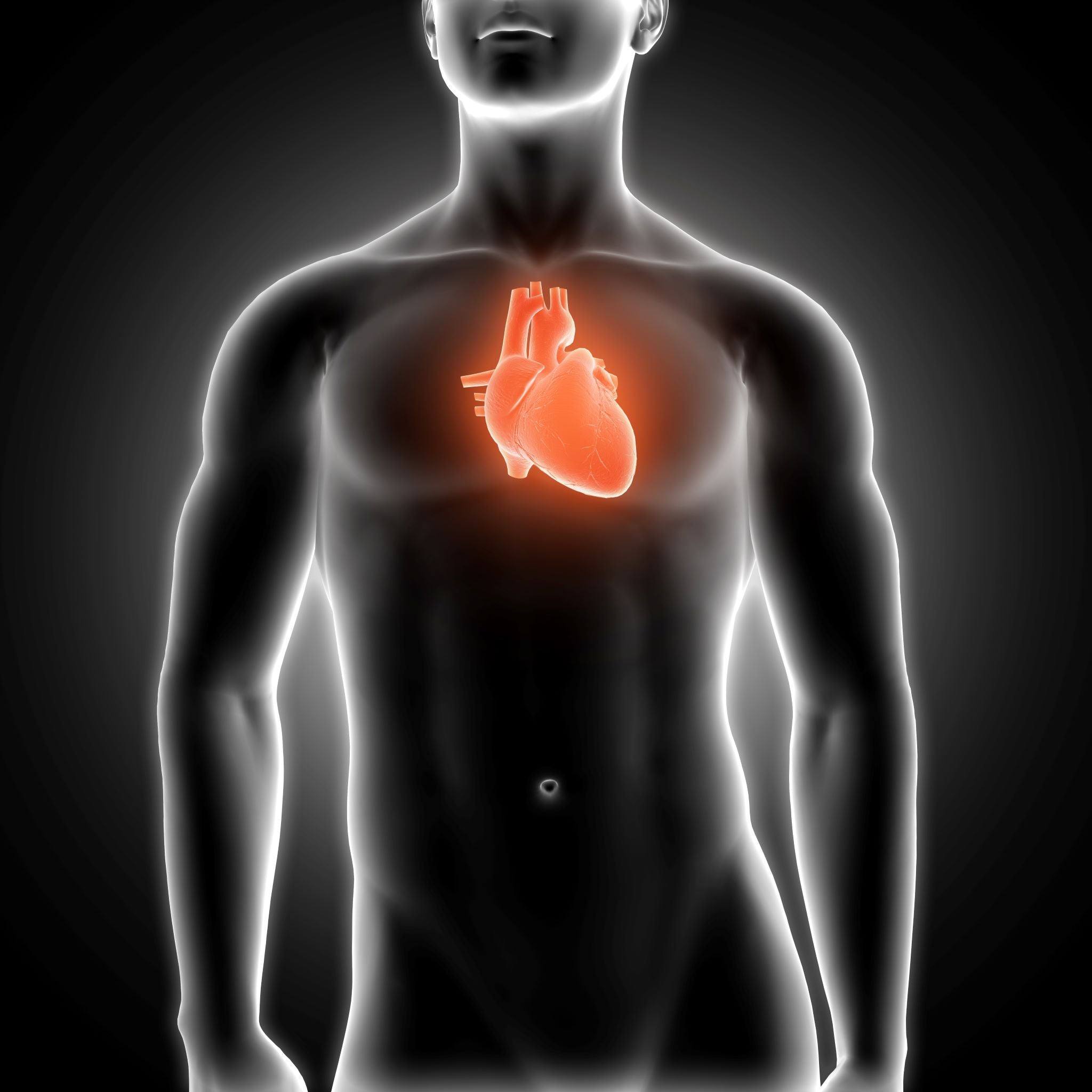
Have you ever experienced the moment when the chest is opened during surgery and the view of the shimmering surface of the heart is revealed? Big as a fist, it pumps blood through your veins full of action and keeps you alive. And in doing so, the heart and its anatomy unfold a fascinating beauty.
You can bring this indescribable view of the surgeon into your home with a drawing from Animus Medicus. You see a frontal view of heart anatomy in accurate detail, just as the surgical team experiences it. With a featherweight of just 300 grams, the heart works around the clock and beats approximately three billion times in our lifetime.
In adults, the healthy heart is up to twelve centimeters long, about eight centimeters thick at its widest point, and six centimeters deep. Protected by the fibrous pericardium, the heart lies in the mediastinum in the center of the chest. On the underside it is firmly connected to the diaphragm. On the right and left it is enclosed by the two lung lobes, against which it is freely movable and separated by the pleura.
Laterally and upward, the largest vessels of the body branch off, which are as thick as a garden hose. The aorta and the pulmonary artery leave the heart. The pulmonary veins and vena cavae flow into the heart.
The vessels and the heart form a closed circulatory system. The left ventricle pumps blood into the aorta. From there it flows into all organs and tissues to supply them with vital oxygen.
The oxygen-poor blood enriched with carbon dioxide flows back into the right atrium, from there into the right ventricle, then into the lungs. There it is enriched with oxygen again and carbon dioxide is exhaled. Then the blood continues into the left atrium and left ventricle, where everything starts over again.
Heart Anatomy: A Muscle That Keeps Us Alive
The largest part of the heart mass is muscle. At first glance at heart anatomy, this appears to be distributed quite simply. The heart is divided into a right and left half, each with a ventricle and an atrium. That makes four heart chambers in total.
But the musculature is much more than a shell: It is built up in several layers that wind in loops and circles from the base of the heart to the apex of the heart.
Through the delicate cooperation of the muscles, the heart manages to contract and eject blood. A drawing like our Heart Overview, whether in vintage look or the black and white Chalk Edition, bathes the beauty of heart anatomy in a very special light. Sit down, take a break and enjoy the elegance of our life pump - while it itself continues its work deep inside you without a break and gives you life.
How fast the heart beats is age-dependent. In adults at rest it is about 60 to 80 beats per minute, in infants about 150. In a healthy person, this pumps the entire blood volume, about five liters in adults, through the body once every minute.
But it's not just human heart anatomy that accomplishes this feat day in and day out! While you can only hear the heart of a mouse ticking at a frequency of 600 per minute, a diving blue whale can eject up to 5000 liters at two beats per minute!
Behind the Scenes: What Does It Look Like Under the Heart Muscle?
The Heart Section drawing available from us even reveals the interior of our miraculous organ. Between the atria and ventricles as well as between the ventricles and the vessels connected to them are the heart valves. The valves are the heart's valves and ensure that blood flows in the right direction and not back where it doesn't belong.
Nevertheless, this can sometimes happen: Heart valve defects and congenital heart defects are among the most common heart diseases. If the valves leak, blood flows back in the wrong direction. If the valves are stuck, calcified or fused, the path for the blood is too narrow. Dizziness and shortness of breath can be the result.
Moreover, valve defects are strenuous for the heart! The musculature can thicken or wear out over time. But even if the heart can change its anatomy, it is still there for you at all times.
While the heart as the body's engine supplies all other organs, its own blood supply is rather inconspicuous. The supply of the heart muscle is ensured by the coronary vessels, also called coronary arteries. They originate directly from the initial section of the aorta and divide into three large branches.
These inconspicuous parts of heart anatomy have a crucial role. If the coronary vessels are narrowed, a heart attack is imminent.
Invisible in our drawing, but indispensable for the complete package, are the nerves of the heart. The pacemaker of the heart is a nerve plexus called the sinoatrial node. It sets the pace and transmits it via nerve cords to the AV node, which transfers the excitation to the ventricles. This way, coordinated information reaches all muscle cells, enabling an orderly beating of the heart muscle.
The nervous system of the heart is even so special that it holds another surprise: It also works all by itself! Even if you were to remove the heart from the body, it could (oxygen and nutrients provided) continue beating on its own for a while!
So if! For now, we have only removed the heart from the body by drawing. So that you can admire the fascinating heart anatomy on your own wall every day.
Share