Spare 50% auf alle Poster

Sort by:
101 products
101 products


The fascination of the fetus:
The development of individual cells is arguably one of the most perfect processes in our lives!
But did you also know the following?
From the 21st week of pregnancy, the fetus can get hiccups. The expectant mother usually perceives this as a slight nudging against the abdominal wall. From the 25th week of pregnancy, voices can already be distinguished from one another, so be careful what you say.
To show how beautiful and exciting pregnancy is, we have created the image Fetus in the Uterus. It is perfect for pregnant women, gynecologists, and midwives.
If you are a doctor, student, medical enthusiast, or nurse, or if you are simply looking for the perfect gift for any occasion, then this artwork is a must-have!
What you get:
- Detailed depiction of the fetus in the uterus in the Chalkboard Edition
- Print on high-quality premium paper
- UV protective laminate for long-lasting colors
- 100% satisfaction guarantee
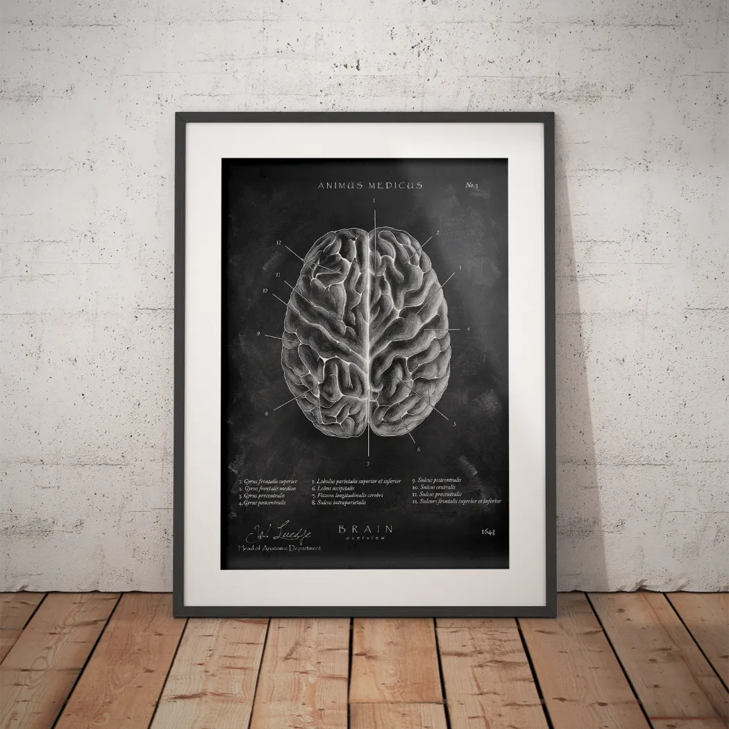
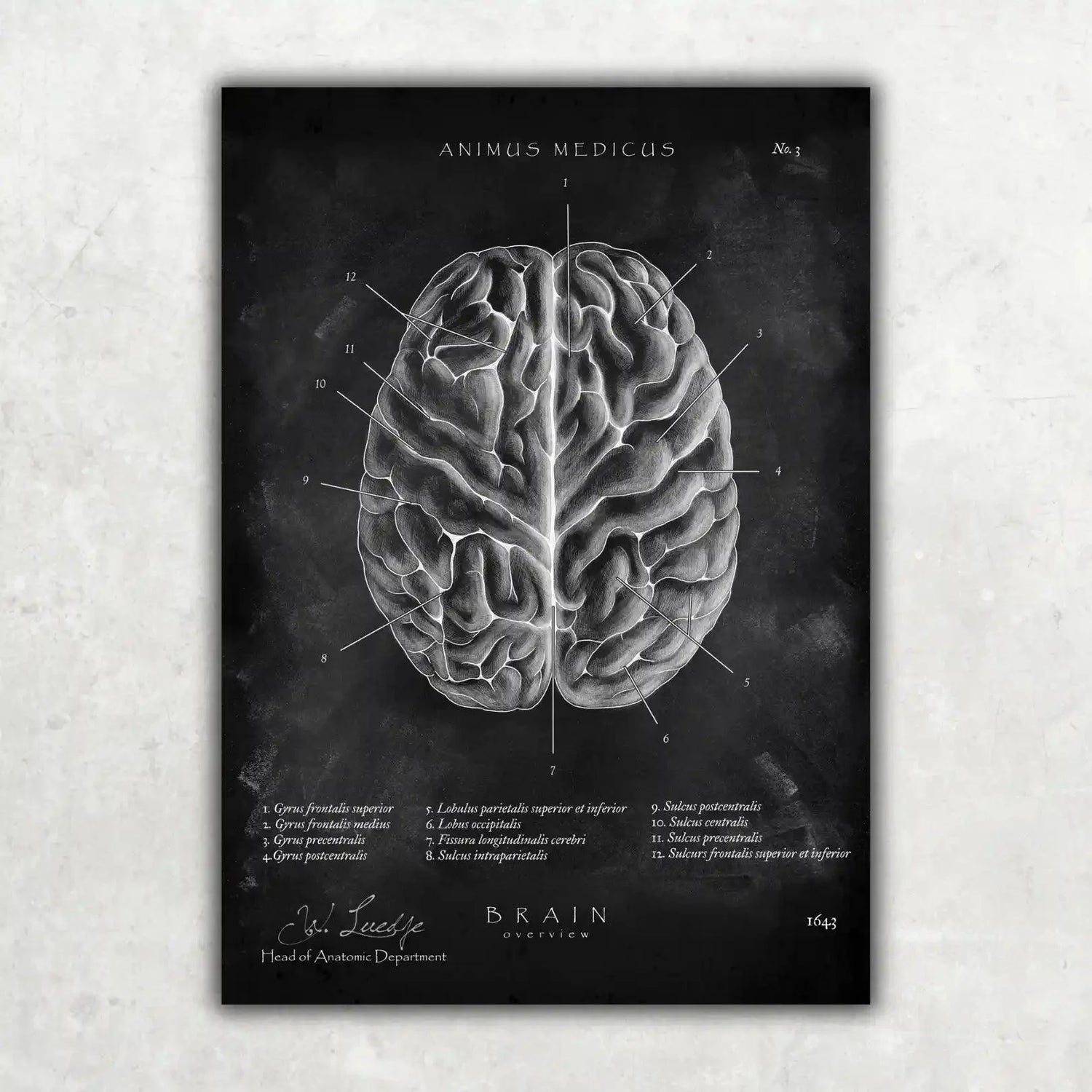
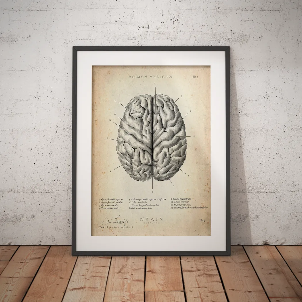
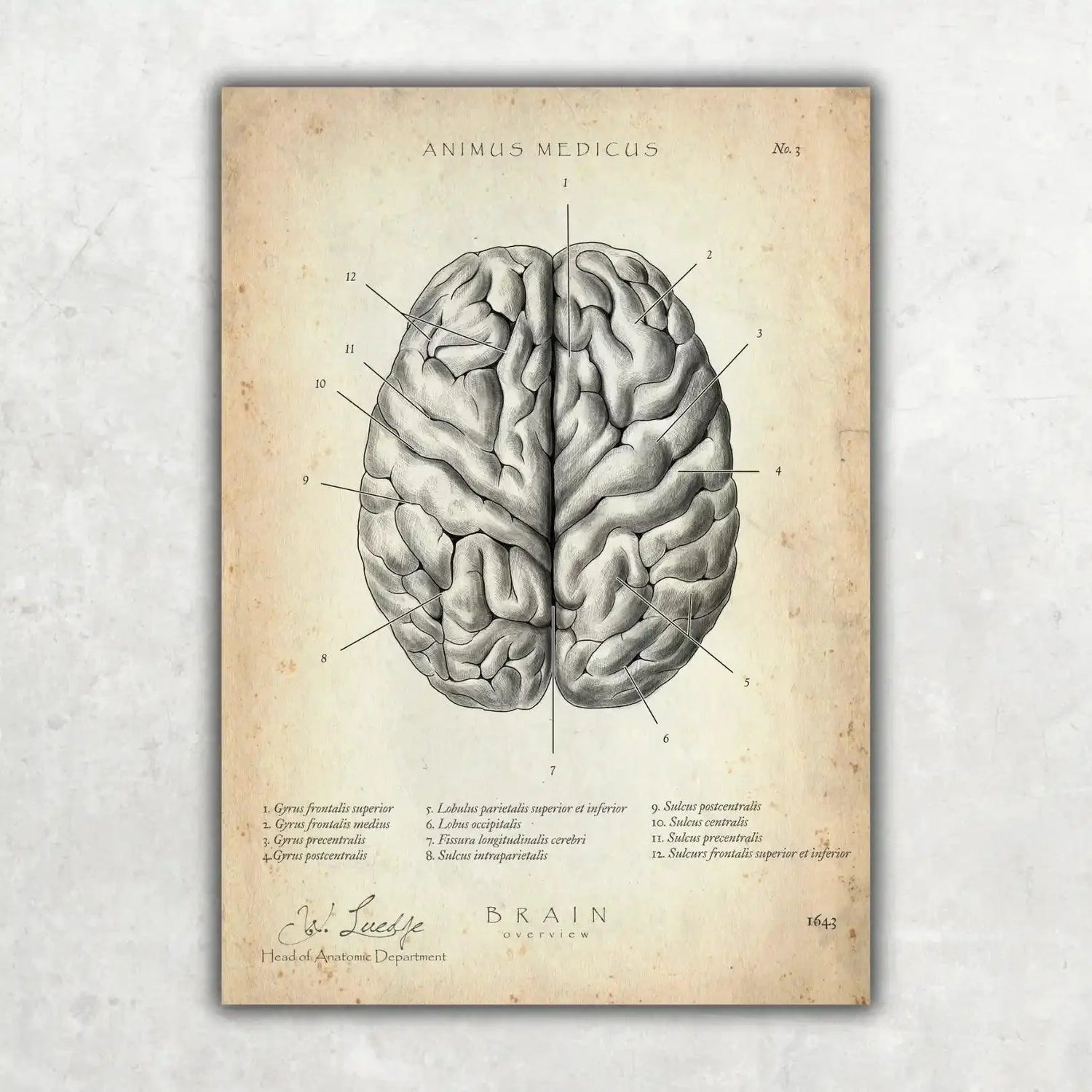
Why the Brain?
The brain is the control center of our body. It makes up only 2-3% of our body weight but consumes up to 20% of the oxygen we breathe in.
Especially in medical fields, having a sharp mind is crucial. Even the smallest mistakes can have significant consequences in medicine.
To ensure that doesn't happen, why not bring another brain home, to your practice, or gift it to someone?
Whether you're a doctor, student, medical enthusiast, nurse, or simply looking for the perfect gift for any occasion, this artwork is a must-have!
What You Get:
- Overview of the brain
- Printed on premium quality paper
- UV-protective laminate for long-lasting colors
- 100% satisfaction guarantee


- Overview of the abdomen
- Printed on high-quality premium paper
- UV protective laminate for long-lasting colors


This poster of the anatomy of the abdomen is perfect for medical practices or your own home and makes an excellent gift for doctors and medical students.
- Overview of the abdomen
- Printed on high-quality premium paper
- UV protective laminate for long-lasting colors
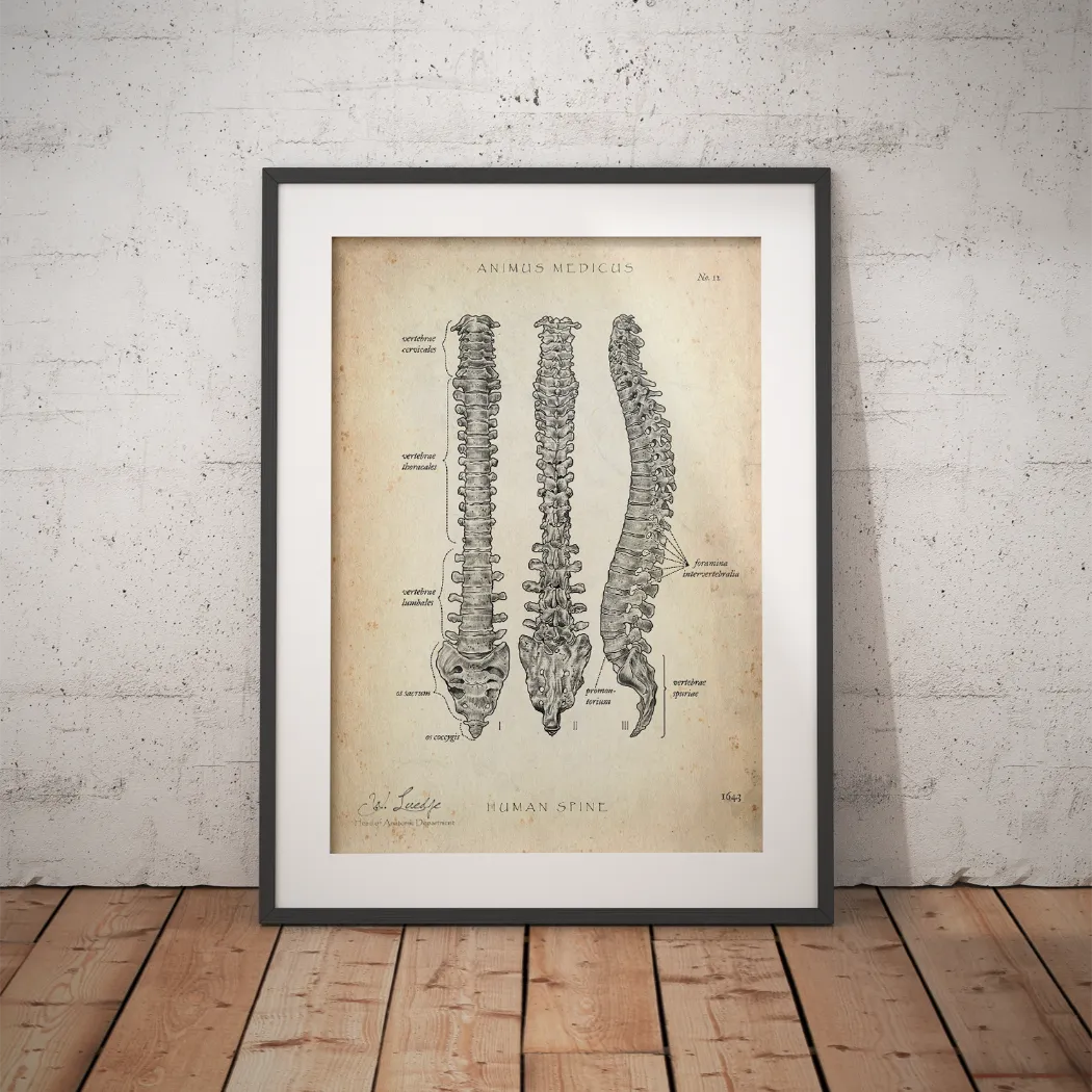

The center of the body
Do you know all the special features of the spine? For most people, their knowledge ends with vertebrae and intervertebral discs. But the spine offers much more than that. ⚕️
It can be divided into 5 sections. 7 cervical vertebrae, the first two of which also have a very special role, 12 thoracic vertebrae, 5 lumbar vertebrae, 5 sacral vertebrae, and 4-5 coccygeal vertebrae.
Explaining this phenomenon to others can quickly become complicated. That's exactly why we created this special motif, which you can use to perfectly explain the spine with its individual sections and special features.
At the same time, it also looks great...
If you are a doctor, student, physical therapist, or nurse, or if you are simply looking for the perfect gift for any occasion, then this artwork is a must-have!
What you get:
- Detailed overview of the spine
- Printed on high-quality premium paper
- UV protective laminate for long-lasting colors
- 100% satisfaction guarantee
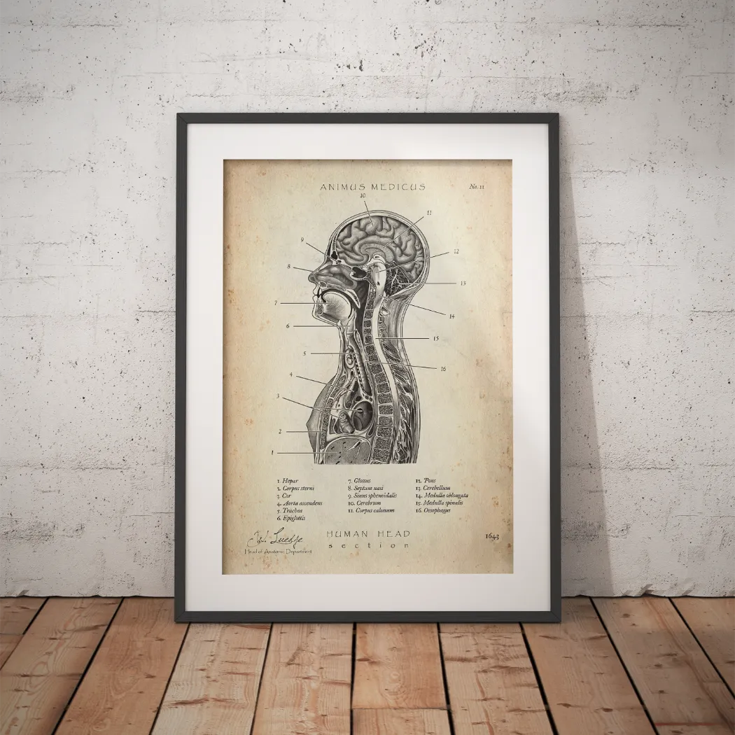
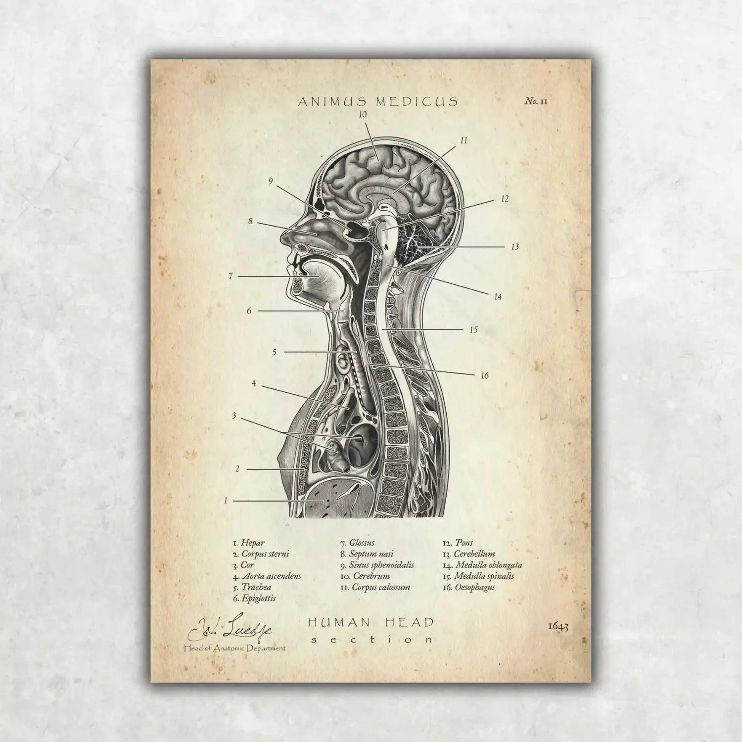
In a sagittal section through the body:
Are you as fascinated by our anatomy as we are? If so, then Head Section is just right for you.
We created this motif to illustrate the numerous organ systems and their location in relation to each other. Now you'll never forget how everything fits together.
And at the same time, it looks incredibly good...
If you are a doctor, student, medical enthusiast, or nurse, or if you are simply looking for the perfect gift for any occasion, then this work of art is a must-have!
What you get:
- Detailed representation of the skull and thorax in sagittal section
- Printed on high-quality premium paper
- UV protective laminate for long-lasting colors
- 100% satisfaction guarantee
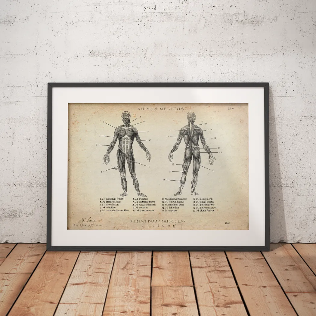
Is the Human Body a Work of Art?
Our answer is a resounding yes! The perfect interplay of muscles, ligaments, bones, and joints is simply impressive.
If you also find the anatomy of the human body fascinating, then you need this poster!
With it, you can admire and study the entire musculoskeletal system of the human body – even outside of anatomy class.
Whether you're a doctor, student, physiotherapist, nurse, or simply looking for the perfect gift for any occasion, this artwork is a must-have!
What You Get:
- Overview of the human musculoskeletal system
- Printed on premium quality paper
- UV-protective laminate for long-lasting colors
- 100% satisfaction guarantee


This art print of the anatomy of the uterus is a wonderful addition to any interior and makes an excellent gift for doctors and medical students.
- Cross-section of the uterus
- Printed on high-quality premium paper
- UV protective laminate for long-lasting colors
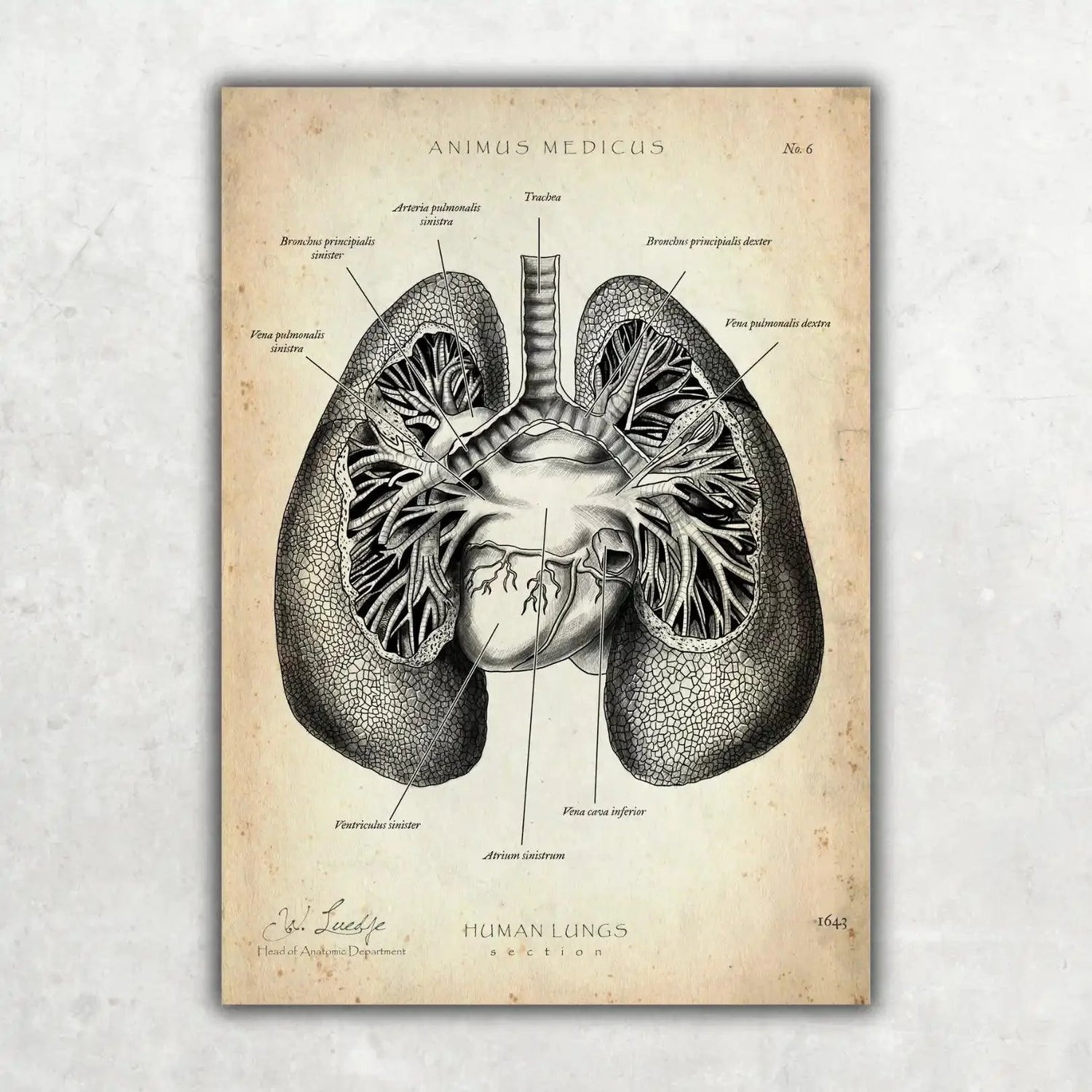
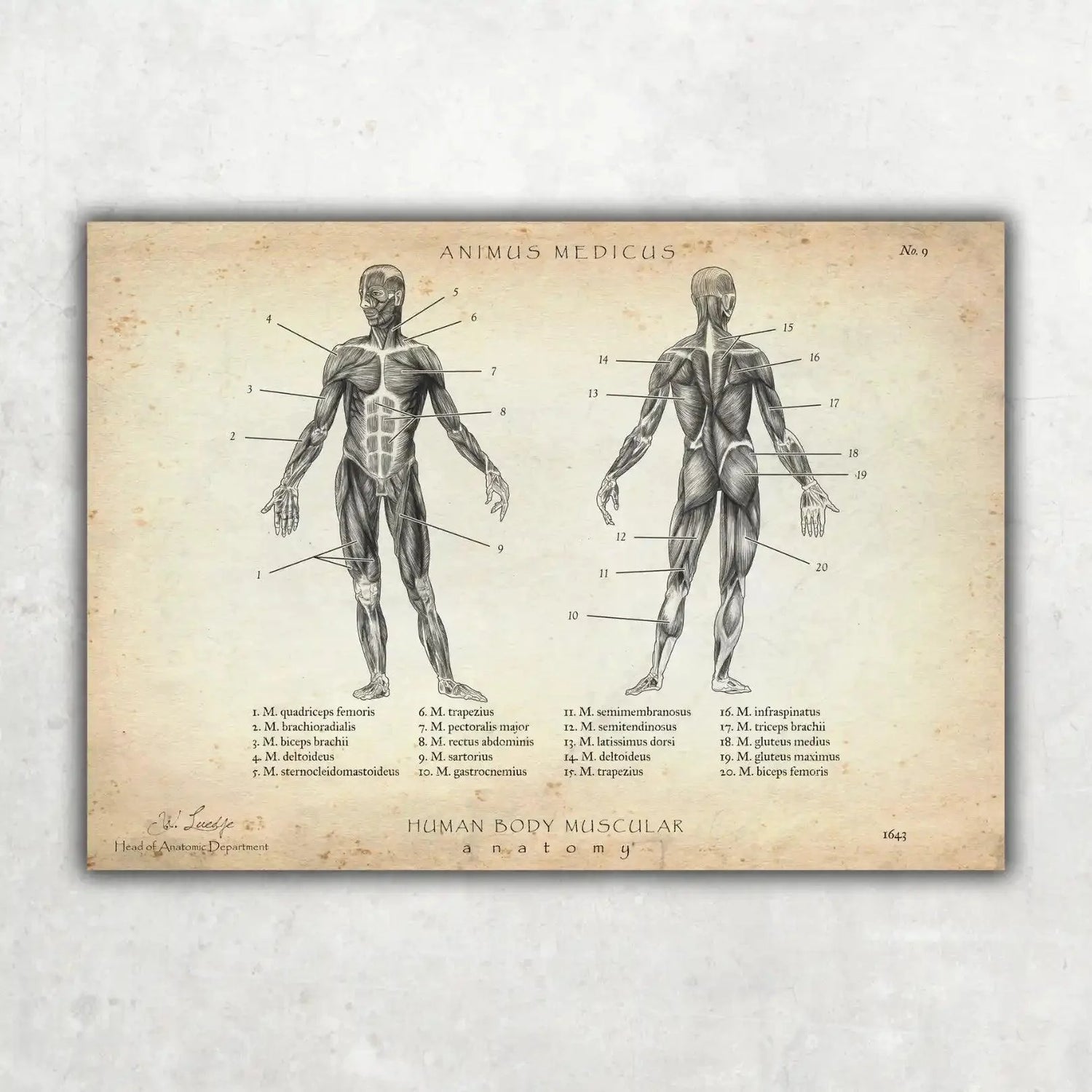
What Makes the Lungs So Special?
Every day, you breathe in up to 20,000 liters of air. This happens with up to 20,000 breaths. Quite impressive, right?
We thought so too. The lungs play a key role in our lives and can help you feel more energetic and relaxed when you use them properly.
Just take 5 deep breaths in and out. With this motif, we remind you to take a moment to breathe deeply and enjoy your life. 🫁
Whether you're a doctor, student, medical enthusiast, nurse, or simply looking for the perfect gift for any occasion, this artwork is a must-have!
What You Get:
- Overview of the lungs
- Printed on premium quality paper
- UV-protective laminate for long-lasting colors
- 100% satisfaction guarantee


- Overview of the teeth
- Printed on high-quality premium paper
- UV protective laminate for long-lasting colors
Why the Brain?
The brain is the control center of our body. It makes up only 2-3% of our body weight but consumes up to 20% of the oxygen we breathe in.
Especially in medical fields, having a sharp mind is crucial. Even the smallest mistakes can have significant consequences in medicine.
To ensure that doesn't happen, why not bring another brain home, to your practice, or gift it to someone?
Whether you're a doctor, student, medical enthusiast, nurse, or simply looking for the perfect gift for any occasion, this artwork is a must-have!
What You Get:
- Overview of the brain
- Printed on premium quality paper
- UV-protective laminate for long-lasting colors
- 100% satisfaction guarantee


The Disc Collection
Explain herniated discs in 60 seconds. Build instant trust.
It's the most common diagnosis—and the most frustrating explanation.
You try your best to explain the difference between a protrusion and a prolapse to your patient.
You talk about nerve roots and spinal canals.
The result? A questioning look and the uncertain question: “So... do I need surgery now?”
Imagine if you could reduce this entire time-consuming process to a single, clear gesture. Pointing to an image that immediately triggers an “aha moment.”
That's exactly why we curated the Disc Collection. Three anatomy posters that form a perfect visual narrative—from healthy overview anatomy to specific pathology.
This is your shortcut to better, faster, and more sustainable patient education.
Your 3-step explanation system:
Step 1: The overview (back anatomy) Show the superficial and deep muscle groups and create a basic understanding of the structure of the back.
Step 2: The zoom (vertebral body anatomy) Go one level deeper and explain the detailed structure of a vertebra and the intervertebral disc as a buffer.
Step 3: The pathology (herniated disc) Visually demonstrate what happens when the buffer gives way and how the pressure on the nerve causes pain.
Your professional advantage:
✔ MAXIMUM CLARITY: Transform complex technical terms into simple, visual truths.
✔ ENORMOUS CONFIDENCE: Position yourself as the expert who takes the time to make problems truly understandable.
✔ HIGHER COMPLIANCE: When patients understand the “why,” their motivation to actually do the prescribed exercises increases.
✔ TIME SAVINGS: Significantly reduce your average explanation time per patient.
“This set has revolutionized the way I talk to back pain patients. Communication is on a whole new level.” - Dr. F. Schmidt, Orthopedist
Join over 3,500 practices and clinics that rely on visual excellence.
Why the Back Anatomy Collection?
✅ Equip your practice with high-quality materials – without compromise.
✅ Create an atmosphere that radiates trust and professionalism.
✅ Impress patients and colleagues at first glance.
✅ Experience daily pride in your modern, inspiring environment.
More than 3,500+ medical facilities already trust in the quality of Animus Medicus.
The set includes:
- 3x posters in your chosen size
- Printed on heavy 200 gsm premium museum-quality paper
- Optional: Add matching premium frames directly
- 30-day satisfaction guarantee & free shipping in the US
Frequently asked questions:
How quickly will my order be delivered?
→ Your set will usually arrive within 4-7 business days.
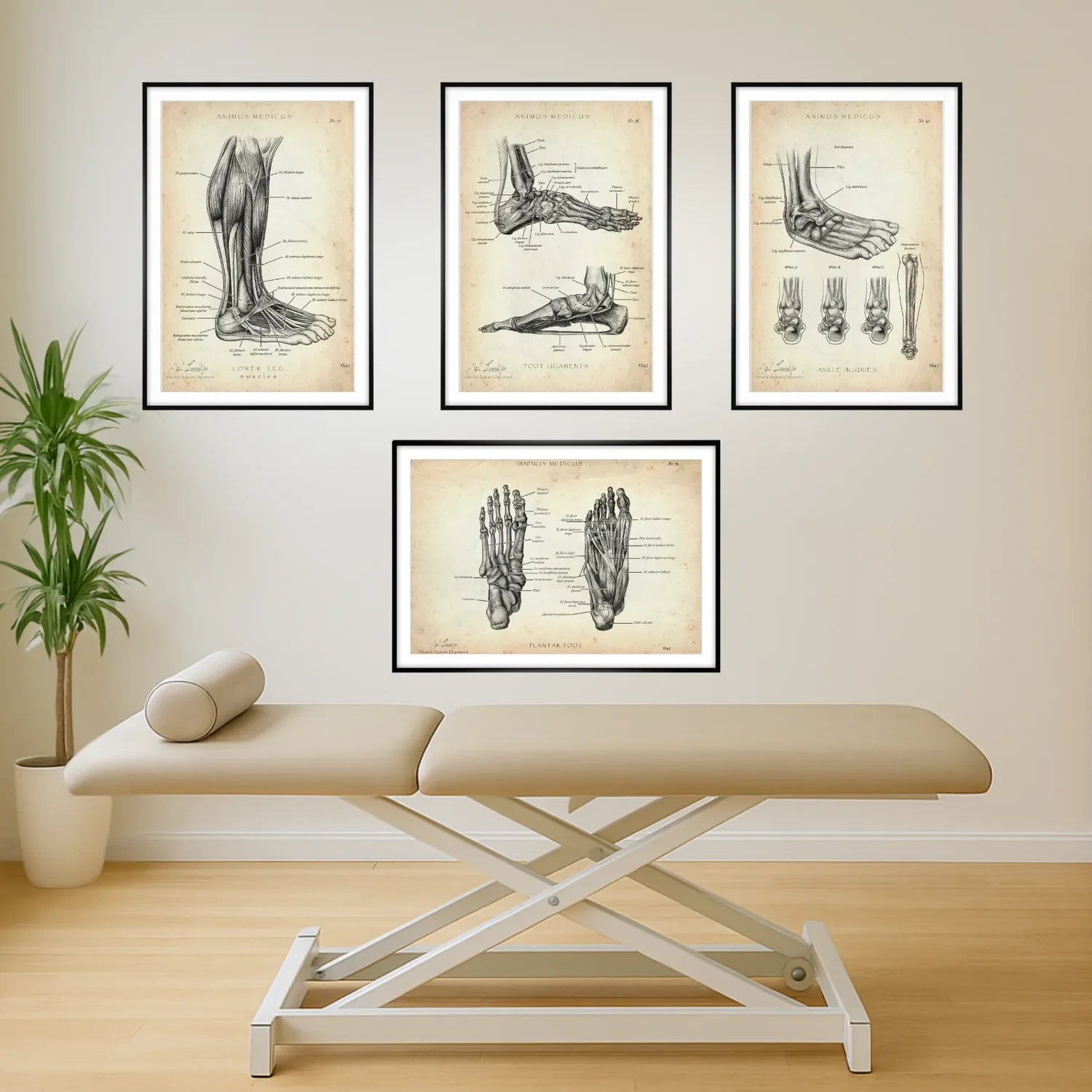

The ankle joint collection for professionals
From “twisting your ankle” to active therapy. Get clarity in seconds.
It's the most frequently asked question after an ankle injury: “When can I start exercising again?”
It's a classic scenario in everyday practice. A patient comes in after a supination trauma—the typical “twisted ankle”—and asks: “It's just a sprain, why do I have to do these weird exercises on the wobble board for 6 weeks?”
You start talking about proprioceptive training and the crucial role of the ligaments in joint stability.
But the patient's motivation for the lengthy training program decreases by the minute.
Imagine if you could show them exactly where the ligaments are overstretched on a poster and visually prove to them why training on the wobble board is the only way to prevent the next sprain.
That's exactly why we've curated this specialized collection. Four teaching aids that illuminate the ankle joint and its surrounding structures from all important perspectives.
It's your visual toolkit for turning passive patients into active, motivated partners.
This is your basis for successful rehabilitation.
Your stability explanation system:
Level 1: The muscles (lower leg muscles) Visualize the muscle chains that actively stabilize the joint and explain the need for strength training.
Level 2: Passive support (foot ligaments) Show the complex ligamentous apparatus and explain what really happens in a “sprain” or torn ligament.
Level 3: The foundation (plantar foot) Demonstrate the crucial role of the arch of the foot for overall statics and the rolling motion.
Stage 4: Trauma (ankle injuries) Clearly explain the mechanics of supination trauma and the classification of fractures such as Weber fractures.
Your professional advantage:
✔ INCREASE THERAPY ADHERENCE: Transform the tedious task of “wobble board exercises” into an understood necessity.
✔ USE TIME EFFICIENTLY: Explain the world's most common sports injury in less than a minute.
✔ REDUCE ANXIETY: Build trust with patients after a traumatic event through visual clarity.
✔ DEMONSTRATE COMPETENCE: Show that you not only treat, but also educate and work preventively at the highest level.
“I use the Ligaments Poster every single day. It has completely changed the way I explain ligament tears. Patients understand it immediately.” - Quote from one of our physio practice customers.
A must-have for any practice focusing on sports medicine, orthopedics, and traumatology.
Join over 3,500 practices and clinics that rely on visual excellence.
Why the Foot Anatomy Collection?
✅ Equip your practice with high-quality materials – without compromise.
✅ Create an atmosphere that radiates trust and professionalism.
✅ Impress patients and colleagues at first glance.
✅ Experience daily pride in your modern, inspiring environment.
More than 3,500+ medical facilities already trust in the quality of Animus Medicus.
The set includes:
- 3x posters in your chosen size
- Printed on heavy 200 gsm premium museum-quality paper
- Optional: Add matching premium aluminum frames directly
-
30-day satisfaction guarantee & free shipping in the US
Frequently asked questions:
How quickly will my order be delivered?
→ Your set will usually arrive within 4-7 business days.

The Shoulder Impingement Collection
From the “painful arc” to a clear treatment plan – in 60 seconds.
It is the most common diagnosis – and the most frustrating explanation.
It is the diagnosis that every therapist knows – and every patient fears. You talk about the “subacromial space,” the “supraspinatus tendon,” and the “painful arc.”
The result? A patient who nods but doesn't really understand why they now have to do tedious external rotation exercises for weeks on end. Compliance drops, frustration rises.
Imagine if you could explain this complex bottleneck with a single, fluid movement of your finger on a poster and create immediate understanding.
This is your visual chain of reasoning for successful therapy.
Your 4-step explanation model:
Step 1: Strength (shoulder muscles) Show the major movers and lay the foundation for the importance of strength training.
Step 2: Mechanics (shoulder joint) Explain the complex interaction of primary and secondary joints.
Step 3: The stabilizer (rotator cuff) Visualize the crucial role of the rotator cuff in joint stability.
Level 4: The problem (shoulder impingement) Demonstrate exactly where and why the tightness occurs and how your therapy increases the space again.
Your professional advantage:
✔ CONVINCE YOUR PATIENTS: Turn passive listeners into active therapy partners.
✔ REFINE YOUR DIAGNOSIS: Use the detailed illustrations for crystal-clear findings.
✔ INCREASE YOUR SUCCESS: Increase your patients' exercise compliance through genuine understanding.
✔ DEMONSTRATE COMPETENCE: Show that you not only treat, but also educate at the highest level.
“Finally, I can show my patients the impingement in a way that they really understand. An absolute game-changer for my daily work.” - Quote from one of our physio practice customers
The first choice for musculoskeletal specialists.
Join over 3,500 practices and clinics that rely on visual excellence.
Why the Shoulder Anatomy Collection?
✅ Equip your practice with high-quality materials – without compromise.
✅ Create an atmosphere that radiates trust and professionalism.
✅ Impress patients and colleagues at first glance.
✅ Experience daily pride in your modern, inspiring environment.
More than 3,500+ medical facilities already trust in the quality of Animus Medicus.
The set includes:
- 4x posters in your chosen size
- Printed on heavy 200 gsm premium museum-quality paper
- Optional: Add matching premium aluminum frames directly
-
30-day satisfaction guarantee & free shipping in the US
Frequently asked questions:
How quickly will my order be delivered?
→ Your set will usually arrive within 4-7 business days.

The knee collection for professionals
Explain cruciate ligament tears, meniscus injuries, and other conditions visually. Save valuable therapy time.
The most frequently asked question after knee surgery is: “When can I start exercising again?”
You begin to explain—about the healing phases of the anterior cruciate ligament, the need to build up the quadriceps, and the danger of putting weight on it too soon.
But all the patient hears is “long break.”
Imagine if you could use a poster to show them exactly why patience is crucial right now and how each of your exercises contributes to the stability of their “new” knee.
That's exactly why we developed this collection.
Four teaching aids that form a complete visual chain: from the basic leg muscles to the crucial stabilizers, to detailed joint structure and the most common injury patterns.
This is the basis for your argument for successful rehabilitation.
Your 4-step explanation model:
Level 1: The foundation (muscles of the leg); Demonstrate the interaction of the entire leg axis and explain why calf and hip training are also important.
Level 2: The powerhouses (upper leg muscles); Focus on the quadriceps and hamstrings – the main drivers of knee stability.
Level 3: The Mechanics (Knee Joint); Explain in detail the structure of cruciate ligaments, collateral ligaments, and menisci.
Level 4: The Trauma (Unhappy Triad); Visualize the classic sports injury and make the complexity of the injury immediately understandable to the patient.
Your professional advantage:
✔ REDUCE CUTTING TIME: Answer complex patient questions in seconds, not in long monologues.
✔ INCREASE COMPLIANCE: Generate genuine motivation for the often lengthy rehabilitation process.
✔EXUDE COMPETENCE: Show your patients (and referring physicians) that you communicate at the highest level.
✔UPSELLING FOR PATIENTS: Visually explain why additional training sessions or kinesio taping are beneficial.
“Since I've had this set, discussions about the duration of rehabilitation have almost disappeared. Patients see it, understand it, and go along with it. Priceless.” - Quote from one of our physio practice customers
Das Standardwerk für jede Praxis mit Schwerpunkt auf Sportverletzungen und post-operativer Reha.
Werde Teil von über 3.500 Praxen & Kliniken, die auf visuelle Exzellenz setzen.
Why the Knee Anatomy Collection?
✅ Equip your practice with high-quality equipment – without compromise
✅ Create an atmosphere that radiates trust and professionalism
✅ Impress patients and colleagues at first glance
✅ Experience pride in your modern, inspiring environment every day
More than 3,500+ medical facilities already trust in the quality of Animus Medicus.
Included in the set:
- 4x posters in your chosen size
- Printed on heavy 200 gsm premium paper in museum quality
- Optional: Add matching premium aluminum frames directly
-
30-day satisfaction guarantee & free shipping
Frequently asked questions:
How quickly will my order be delivered?
→ Your set will usually arrive within 4-7 business days.


Balance & Hearing — Beautifully Visualized
Ever felt dizzy after a roller coaster or had ringing ears after a concert? Then you're already well acquainted with your inner ear.
This poster helps you understand the structure of the cochlea and vestibular organ — and appreciate their extraordinary complexity. Physician-designed for accuracy, stunning enough to hang in any clinic or office.
What You Get:
- Inner Ear Anatomy poster
- Printed on premium heavyweight paper (200 g/m²)
- UV-protective laminate for long-lasting colors
- 100% satisfaction guarantee
Trusted by 3,500+ clinics and practices worldwide.
Showing 100/101
- Satisfaction guarantee
- Fast delivery
- Buyer protection
- Satisfaction guarantee
- Fast delivery
- Buyer protection