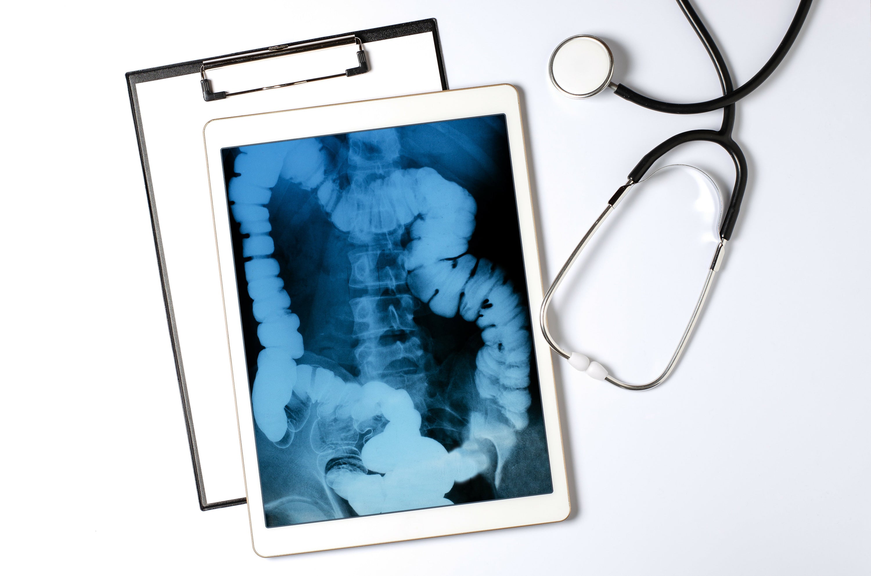
X-rays Explained: A Fascinating Look Inside
Ever wondered what lies beneath your skin?
X-rays can reveal just that. During an X-ray examination, a specially trained medical professional can see exactly what your bone structure looks like and interpret what this imaging reveals. The discovery of X-rays by Wilhelm Conrad Röntgen in 1895 was a milestone that satisfied the prevailing curiosity about the interior of the human body – it allows us to literally look beneath the skin. This article by Animus Medicus brings you closer to the world of X-rays and explains what to expect during an X-ray examination. This way, you can approach your next X-ray examination with ease and learn some fascinating things about X-ray technology.
What are X-rays?
X-rays are a form of electromagnetic radiation, similar to the light we see or the radio waves that transmit music and news. The difference lies in their wavelength: X-rays are much shorter, which gives them the ability to penetrate certain materials, such as our bodies.
X-rays were discovered accidentally by the physicist Wilhelm Conrad Röntgen, who noticed a mysterious radiation that darkened photographic plates while experimenting with cathode ray tubes. Röntgen's discovery revolutionized physics on one hand and became an indispensable tool in medical diagnostics on the other.
How an X-ray machine works
The function of an X-ray machine is to create an image of something that lies beneath an opaque surface. In a classic medical X-ray machine, it essentially involves a tube that shoots electrons at a piece of metal. When the electrons hit, X-rays are produced and passed through the body being examined.
Since different materials absorb the rays differently, an image is created on a detector or photographic plate. Bones appear white, soft tissues in shades of gray, and air appears black. Nowadays, digital X-ray techniques can directly convert these rays into images displayed on a screen. This modernization allows for immediate assessment of the imaging by the medical professional.
X-ray explained: The procedure
For those who have never experienced an X-ray examination, it might be hard to imagine. Here’s how an X-ray examination typically proceeds:
-
Preparation:
- When it's your turn for the X-ray, you are often first taken to a regular treatment room before entering the room with the X-ray machine. The X-ray procedure is often briefly explained to you.
- You may be asked to remove certain clothing items, such as medical socks for foot injuries, and change into a gown. Personal items like anatomical jewelry and piercings should also be removed before the X-ray.
-
Safety Measures:
- Although X-rays in high doses can be harmful, the amounts used in a typical X-ray examination are very low. Nevertheless, lead aprons or other protective measures are used to shield body parts that are not being examined.
-
The X-ray:
- An X-ray is quick and painless. You will be asked to stand or lie still while the X-ray machine is briefly activated. Your position depends on the body part being imaged. The medical staff will help you position yourself to obtain the best possible image. It is crucial to stay still during the brief moment the image is taken to ensure clarity.
-
After the X-ray:
- Once the images are taken, you can get dressed and leave the room. The entire procedure often takes only a few minutes, and the results are evaluated by a radiologist, who discusses the findings with you and your treating physician.
Understanding anatomy through X-rays of bones
What’s remarkable about X-rays is how they reveal the hidden structures of our bodies. They enable doctors to diagnose fractures, locate foreign objects, or assess the health of joints. X-rays are not limited to the human body; they also provide insights into the anatomy of animals, which is essential for veterinary diagnoses. X-ray imaging fascinates not only medical professionals but also the general public, as it makes visible what is invisible to the naked eye.
The importance of X-rays for diagnosis
X-rays are often the first step in the diagnostic process. Without them, a fracture might only be guessed. The anatomical images from an X-ray can reveal conditions that are not detectable through external examination methods. These include the presence of tumors, the extent of an infection, the presence of a bone injury, or the stage of a disease. Interpreting X-ray images requires expertise, as anomalies in bones and other parts of the human anatomy are not always easy to recognize and interpret. Your specialist will explain your X-ray results in detail and recommend further treatment.
X-rays beyond medicine
The application of X-rays is not limited to the medical field. Here are some other fascinating areas where X-rays are used:
-
Art and Archaeology:
- Analysis of artworks and archaeological finds, revealing hidden layers and previous modifications, and estimating the age of bones based on their condition.
-
Industry:
- Examination of material structures and detection of flaws. X-rays can identify cracks or air pockets, allowing for the sorting out of defective materials.
-
Security Technology:
- Detection and prevention of hazards, such as at airports. Objects and clothing are scanned for dangerous items.
-
Other Fields:
- Geology, astronomy, and many other areas also benefit from X-ray technology.
The indispensable technology of X-rays
X-ray machines and their function allow us to gain deep insights into the world around us and within us. This technology has revolutionized medical diagnostics and finds applications in many other areas.
Interested in anatomy and medicine?
Did you enjoy this article and are interested in anatomy and medicine? Then perhaps our anatomy phone cases or anatomy pins from Animus Medicus are something for you. Visit our shop and discover our unique products!
Share